| Introduction to the technique | Pictures of our AFM instrument |
| Tip 'Convolution' in SPM | Mark's AFM image gallery |
| Links to AFM and SPM Tutorials |
Introduction to AFM
AFM is one of many techniques which fall under the Scanned Probe Microscopy (SPM) family of instruments. In all of these SPM techniques a small probe (10-100 nm radius of curvature) is raster scanned by a piezoelectric device over a sample to produce an image of the sample surface, or near surface region.
The first of the SPM techniques was the Scanning Tunneling Microscope (STM), developed by Binnig & Rohrer, which got them the Nobel prize for Physics in 1986. The contrast mechanism in the STM is based on the tunneling of electrons from a sharp metal probe tip to a conductive sample.
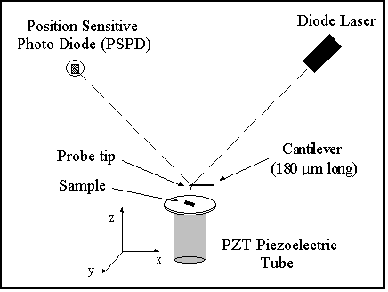
A simple schematic of an AFM instrument is given in Figure 1, below. In this instrument the probe tip is mounted on the end of a triangular cantilever arm, similar to a diamond stylus mounted on the end of a record player arm. A piezoelectric device raster scans the sample beneath the probe tip. As the probe tip undergoes attractive or repulsive forces, the cantilever will bend. This bending of the cantilever can be monitored by bouncing a laser beam (a simple diode laser, like that found in a CD player works well) off of the cantilever onto a 2 element photodiode (Position Sensitive Photo Diode). In normal operation the tip-sample force is held constant by a computer controlled feedback loop that examines the force (bend of the cantilever) and tells the piezoelectric device whether to move the sample closer or farther away in order to maintain the set force value. The AFM image produced by taking the feedback signal at each pixel of the raster scan, is a measure of the topography of the sample.
Figure 1: AFM schematic
The AFM uses the attractive or repulsive forces
encountered by a probe tip when it is in close proximity to a sample surface <200 nm). There are three main modes of AFM operation that are currently in use:
Contact, Non-Contact and Intermittant Contact (Tapping). Contact AFM is done by bringing
the tip to a distance at which repulsive forces dominate the tip-sample interaction.
Non-Contact AFM is done such that the tip-sample interaction is in the attractive or van
der Waals regime. In order to perform measurements in this attractive force region the
cantilever is oscillated with a low amplitude (<5 nm), near its resonant frequency. For
Non-Contact AFM the force is measued by comparing the frequency and/or amplitude of the
cantilever oscillation relative to the driving signal. Tapping or Intermittant Contact
mode is also done by oscillating the cantilever near its resonant frequency, but the
amplitude is significantly higher (~10-50 nm?). This Intermittant contact mode operates in
the repulsive force region, but touches the surface only for short periods of time, in
order to reduce damage to potentially fragile sample (ie. biological molecules).
(<200 nm). There are three main modes of AFM operation that are currently in use: Contact, Non-Contact and Intermittant Contact (Tapping). Contact AFM is done by bringing the tip to a distance at which repulsive forces dominate the tip-sample interaction. Non-Contact AFM is done such that the tip-sample interaction is in the attractive or van der Waals regime. In order to perform measurements in this attractive force region the cantilever is oscillated with a low amplitude (<5 nm), near its resonant frequency. For Non-Contact AFM the force is measued by comparing the frequency and/or amplitude of the cantilever oscillation relative to the driving signal. Tapping or Intermittant Contact mode is also done by oscillating the cantilever near its resonant frequency, but the amplitude is significantly higher (~10-50 nm?). This Intermittant contact mode operates in the repulsive force region, but touches the surface only for short periods of time, in order to reduce damage to potentially fragile samples (ie. biological molecules).
The finite size and the shape of the probe tip lead to imaging artifacts, such as tip 'convolution', which normally limits the lateral resolution to about 5-10 nm.
For some good tutorial information on Scanning Probe Microscopy try the following sites:
visits since 2/11/00