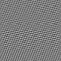
This is illustrated by the AFM image of mica above, which is approximately 225 Å x 225 Å.
Probe Tip 'Convolution' in Scanning Probe Microscopes
Crystalline samples, cleaved along
atomically flat planes can be imaged with lattice and atomic resolution.
This is illustrated by the AFM image of mica above, which is approximately
225 Å x 225 Å.
Most samples, however, have features that are on the same size scale as the probe tip used for imaging, which 'convolute' with the size and shape of the probe tip during the imaging process. This is shown in the animation below.

This animation shows a model of a probe tip scanning over a flat surface which has a spherical particle adsorbed on it. The black line is the data that the AFM records. The result of this convolution effect is illustrated below for a real sample.
Below is an image of Tobacco Mosaic Virus (TMV) adsorbed to a mica substrate.
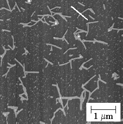
The TMV is a cylinder with diameter of 180 Å. It is clear from a cross section through the above image (indicated by the white bar) that the height is accurately measured to be ~180 Å, but the width is overestimated by nearly a factor of two!
During my dissertation work I helped to develop a way around this problem for the measurement of protein structure at the solid/liquid interface. I should get more information posted about this technique and my interests in the field soon! Drop me an email if you are interested in more information.