 .
.AFM Image Gallery
 .
.
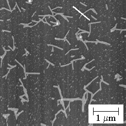
AFM image of Tobacco Mosaic Virus on mica.

AFM image of Streptavidin layer on mica. Note that the center region, which has been
scraped away, was scanned at a high repulsive force.

Line scan from above image of Streptavidin layer on mica.
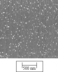
AFM image of individual Concanavalin A molecules on a mica surface.
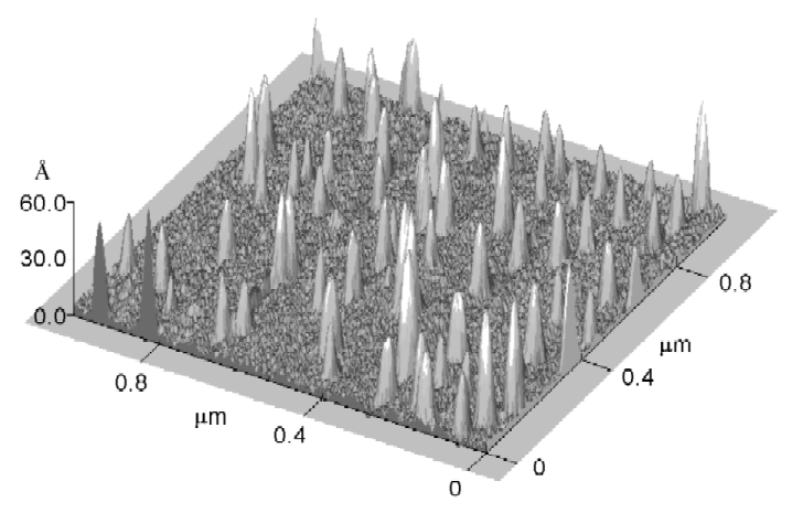
Three dimensional rendering of Concanavalin A on mica.
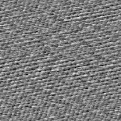
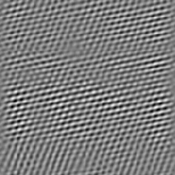
Unfiltered (Left) and Fast Fourier Transform (FFT) filtered (Right) AFM image of freshly
cleaved mica, illustrating lattice scale resolution of AFM.

AFM image of a yeast nuclear pore complex
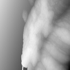
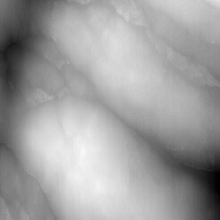
5 mm (Left) and 2 mm (Right) image
of Lilly pollen.

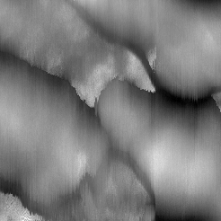
1 mm (Left) and 0.5 mm (Right) image
of Lilly pollen.